
DigiPatICS: Projecte d'optimització del diagnòstic anatomopatòleg en xarxa als hospitals de l'Institut Català de la Salut a través de la digitalització i eines d'intel·ligència Artificial
| Type | Start | End |
|---|---|---|
| Other | Oct 2020 | Dec 2026 |
| Responsible | URL |
|---|---|
| Ferran Marqués & Philippe Salembier | DigiPatICS notice page @ICS |
Reference
Institut Català de la Salut (ICS). Palex Medical. Programa Operatiu FEDER de Catalunya 2014-2020.
Description
GPI collaborates with the Institut Català de la Salut (ICS) in the context of the DigiPatICS project. The first phase of DigiPatICS (10/2020-12/2023) was conducted under the Framework of the Catalonia FEDER Program where UPC is an LTP of Palex Medical. The second phase (08/2024-12/2025) is directly funded by ICS with a potential extension until 2027.
DigiPatICS aims to enhance patient safety and quality of care by improving the diagnosis of histopathological samples and optimizing ICS digital pathology (DP) services and Artificial Intelligence (AI) tools.
These tools have been deployed in 8 ICS hospitals: HU Bellvitge, HU Doctor Trueta, H. Verge de la Cinta, HU Germans Trias i Pujol, HU Joan XXIII, Viladecans Hospital, HU Arnau de Vilanova, and HU Vall d'Hebron. It involves over 170 pathologists, 24 scanners connected to 13 macropath stations, generating a million scanned images annually (Whole Slide Images-WSI). Possibly one of the largests DP networks in the world.
 |
 |
| DigiPatICS scanners installed in ICS Hospitals. Source: ICS news 03/2021 |
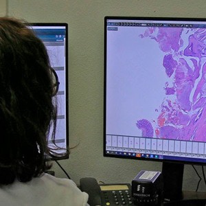
Pathologist and resident diagnosing in the office. Source: Temprana-Salvador et al (2022) |
GPI has developed algorithms to assist pathologists’ diagnosis by automatically analyzing WSI images, enabling rapid and consistent biomarker quantification and statistically robust results. Initially, ICS focused the project on breast cancer due to its high prevalence (it is estimated that one in eight women has had or will have breast cancer). Currently, we develop new algorithms to support doctors in other pathologies: analysis of Hematoxylin & Eosin (H&E) images for breast and lung cancer, in-situ hybridization (ISH) for gastric cancer, cytology for bronchoalveolar lavage (BAL) studies, and radiographic images for knee osteoarthritis assessment.
In a biopsy for suspected breast cancer, a tissue section is first analyzed using H&E staining to detect cancer presence. Other sections are immunohistochemically stained (IHC) to identify the cancer type by computing cells reacting to biomarkers and determining the optimal treatment.
Notably, WSI images are enormous, in the order of tens of GigaPixels (100Kpix x 200Kpix) and contain tens of thousands cells. Manual counting by pathologists on small tissue areas (about a hundred cells each) is assumed to be representative, but may lose statistical significance.
GPI has created deep learning models to study, detect, and classify cells in IHC stains for pathologists' diagnoses. Semantic and instance segmentation algorithms with convolutional and graph-based neural networks are tailored to each medical problem. These algorithms process all cells in WSI images offline (e.g., overnight), and results are stored for quick access. Pathologists visually supervise the result and retrieve calculated values to quantify biomarkers over selected areas, often much larger than those chosen for manual calculation, improving statistical precision of diagnosis.
In the first stage of the DigiPatICS project, four algorithms were developed for analyzing stains based on the presence of HER2 and Ki67 proteins, and estrogen and progesterone receptors.
Students' contributions to DigiPatICS
By project type and in descending order of submission date.
PhD Thesis
- Pina Benages, Òscar (2025, June 30). From pixels to graphs: cell-based digital pathology image analysis, PhD Thesis, Signal Theory and Communications, UPC, adv. V. Vilaplana
Master Thesis
- González Mestre, Adrià (2025, May 27). Breast tumor segmentation in histological Whole-Slide Images using nnU-Net (tmplink), Master Thesis MATT, ETSETB-UPC, adv. V. Vilaplana, L. Jiménez
- Sánchez Bergés, Marc (2024, July 11). Automatic registration of breast cancer tissue, Master Thesis MATT, ETSETB-UPC, adv. V. Vilaplana
- Rabanaque Rodríguez, Sonia (2024, January 22). Immunohistochemistry images analysis with convolutional and graph neural networks, Master Thesis MAI, FIB-UPC/URV/UB, adv. P. Salembier, J.R. Casas
- Lin Huang, Peng (2023, July 5). U-Net vs HoVer-Net: A Comparative Study of Deep Learning Models for Cell Nuclei Segmentation and Classification in Breast Cancer Diagnosis, Master Thesis MATT, ETSETB-UPC, adv. P. Salembier, F. Marqués
- Cavallari, Lucia (2022, July 12). Non-rigid registration on histopathological breast cancer images using deep learning, Master Thesis MET, ETSETB-UPC, adv. V. Vilaplana, J.R. Casas
- Espina i Boronat, Maria (2022, July 5). Analysis and segmentation of KI-67 immunohistochemistry images for breast cancer diagnosis, Master Thesis MATT, ETSETB-UPC, adv. P. Salembier, M. Pardàs
Degree Project
- Jansat Ballarin, Berta (2024, October 29). Cell segmentation and quantification with multi-task approach including uncertainty estimation, Bachelor Thesis GRETST, ETSETB-UPC, adv. M. Pardàs, D. Anglada
- Sáez i Parés, Laura (2024, October 23). Analysis of histhopatological images for assessing cancer samples, Bachelor Thesis DSE, ETSETB/FIB-UPC, adv. F. Marqués
- Fortuño Martí, Héctor (2024, June 28). Analysis of immunohistochemistry images with Hematoxylin and Eosin staining for lung cancer diagnosis, Bachelor Thesis DSE, ETSETB/FIB-UPC, adv. P. Salembier
- Caro Via, Valèria (2024, June 27). Selection and integration of H&E algorithms in the DigiPatICS pipeline, Bachelor Thesis DSE, ETSETB/FIB-UPC, adv. J.R. Casas, V. Vilaplana
- Qiu, Yikai (2023, June 29). Breast Cancer Tissue Classification with Contrastive Learning on Whole Slide Images, Bachelor Thesis DSE, ETSETB/FIB-TSC-UPC, adv. J.R. Casas, S. Rabanaque
- Pérez Cano, José (2023, June). Automated detection of tumoural cells with graph neural networks, Bachelor Thesis MATH/DSE, CFIS/ETSETB/FIB-TSC-UPC, adv. P. Salembier, F. Marqués
- Segon Mayans, Maria Elisabet (2023, January 26). Histopathology Image Analysis for Breast Cancer Diagnosis, Bachelor Thesis DSE, ETSETB/FIB-TSC-UPC, adv. P. Salembier, F. Marqués
- Rosell Murillo, Marina (2022, October 19). Segmentation and classification of tumor cells in breast cancer histological images: analysis of multicenter variability, Bachelor Thesis DSE, ETSETB/FIB-TSC-UPC, adv. F. Marqués, M. Pardàs
- Blanco i Arnaus, Júlia-Ariadna (2022, July 5). Detection and quantization of biopsy samples from biopsy cassette images, Bachelor Thesis GRETST, ETSETB-UPC, adv. F. Marqués, J.R. Casas
- Gaston Codony, Fernando (2022, June 27). Tumorous area detection in breast cancer biopsy images using multi-resolution networks, Bachelor Thesis DSE, ETSETB/FIB-UPC, adv. V. Vilaplana, J.R. Casas
- Pereira Cánovas, Anxo-Lois (2022, June 27). Colon nuclei histological images segmentation and classification, Bachelor Thesis DSE, ETSETB/FIB-UPC, adv. F. Marqués, M. Pardàs
- Campos Almazán, Alejandro (2021, July 5). BAL images analysis for their automatic quantification, Bachelor Thesis GRETST, ETSETB-UPC, adv. F. Marqués, M. Pardàs
- Boneta Camí, Mireia (2021, June 28). Analysis of HER2 receptor proteins in breast cancer histology images using semantic segmentation, Bachelor Thesis DSE, ETSETB/FIB-UPC, adv. M. Pardàs, P. Salembier
- Magariño Llavero, Pau (2021, June 28). Deep Learning based segmentation and classification of stained nuclei breast cancer histology images, Bachelor Thesis DSE, ETSETB/FIB-UPC, adv. F. Marqués, M. Pardàs
- Rabanaque Rodríguez, Sonia (2021, June 28). Breast tumor detection in Hematoxylin & Eosin stained biopsy images, Bachelor Thesis DSE, ETSETB/FIB-UPC, adv. J.R. Casas, V. Vilaplana
- Sala Prat, Júlia (2021, June 28). Cell detection and classification of breast cancer histology images using a deep learning approach based on the U-Net architecture, Bachelor Thesis DSE, ETSETB/FIB-TSC-UPC, adv. P. Salembier, M. Pardàs
i2Rs/CBIs/internships
- Sáez Parés, Laura (Spring 2024). Development of biomedical analysis algorithms, Internship report, DSE degree, FIB/ETSETB-UPC, adv. F. Marqués
- Beaus Iranzo, Pablo (Spring 2023). Tissue Detection in Whole Slide Images, i2R report, MATT master, ETSETB-UPC, adv. S. Rabanaque, J.R. Casas, F. Marqués
- Le Boeuf Flo, Andres (Spring 2023). Detection and classification of cancerous cells, Internship, MATT master, ETSETB-UPC, adv. M. Pardàs, F. Marqués
- Rodríguez Muñoz, Martín (Spring 2023). Analysis and segmentation of immunohistochemistry images for breast cancer diagnosis: HER2 Staining, I2RCED report, DSE degree, FIB/ETSETB-UPC, adv. P. Salembier, F. Marqués
- Qiu, Yikai (Fall 2022). Ductal Carcinoma in situ (DCIS) segmentation on HE whole slides by... truth, I2RCED report, DSE degree, FIB/ETSETB-UPC, adv. F. Marqués, J.R. Casas, S. Rabanaque
- Rubio Jornet, Víctor (Fall 2022). Improving WSI registration for cross-slide histopathological analysis of multiple stains, i2R report, MATT master, ETSETB-UPC, adv. J.R. Casas, S. Rabanaque
- Formosa Marin, Feliu (Spring 2022). Cell detection and classification of lung cancer histological images using 2 single-purposed parallel neural networks, i2R report, MATT master, ETSETB-UPC, adv. P. Salembier.
- González i Juclà, Daniel (Fall 2021). Cell nuclei detection and segmentation from registered HER2 and CK19 histological images, I2RCED report, DSE degree, FIB/ETSETB-UPC, adv. J.R. Casas, V. Vilaplana
- Pereira Cánovas, Anxo-Lois (Fall 2021). Cancerous nuclei segmentation and classification in histological images models comparison, I2RCED report, DSE degree, FIB/ETSETB-UPC, adv. F. Marqués, P. Salembier, M. Pardàs
- Turch Ferreres, Arnau (Fall 2021). Upgrading the breast tumor detection in Hematoxylin & Eosin stained biopsy images, I2RCED report, DSE degree, FIB/ETSETB-UPC, adv. J.R. Casas, V. Vilaplana
- Pereira Dos Santos, Victor (Spring 2021). In-situ tumor detection in H&E images, Internship, MATT master, ETSETB-UPC, adv. M. Pardàs, F. Marqués
- Pujol Salido, David (Spring 2021). Tumor detection in HER2 images, I2R report, MATT master, ETSETB-UPC, adv. V. Vilaplana
- Rabanaque Rodríguez, Sonia (Spring 2021). Alignment of multi-stained digital histological images, CBI report, DSE degree, FIB/ETSETB-UPC, adv. J.R. Casas, V. Vilaplana
- Ramírez Márquez, Alex (Spring 2021). Tumor detection in HER2 images, I2R report, MATT master, ETSETB-UPC, adv. V. Vilaplana
- Aguilera González, Cristina (Fall 2020). Cell nuclei detection and segmentation in histological images based on a deep semantic segmentation network, CBI report, DSE degree, FIB/ETSETB-UPC, adv. F. Marqués, P. Salembier
- Fernández López, Christian (Fall 2020). HER2 nuclei and membrane segmentation, I2R report, MATT master, ETSETB-UPC, adv. V. Vilaplana
- Magariño Llavero, Pau (Fall 2020). Cancerous nuclei segmentation using object detection, CBI report, DSE degree, FIB/ETSETB-UPC, adv. F. Marqués, M. Pardàs
- Rosell Murillo, Marina (Fall 2020). Use of Image 2 Image translation to handle variations between multicentric HER2 stains of breast cancer biopsy, CBI report, DSE degree, FIB/ETSETB-UPC, adv. F. Marqués, M. Pardàs
Current students (MsTh, DegProj, i2R/CBI/internships)
- Alonso del Hoyo, Sara (exp. Spring 2025). Automatic quantization of Bronchoalveolar Lavage (BAL) images, Bachelor Thesis DSE, ETSETB/FIB-UPC, adv. M. Pardàs, F. Marqués
- Caus Solé, Albert (Spring 2025). Co-registration of histopathological images in the DigiPatICS project, internship and Bachelor Thesis GRETST, ETSETB-UPC, adv. J.R. Casas
- Ramon Montmany, Laura (Spring 2025). Breast tumor detection in histological images using deep learning, Bachelor Thesis DSE, ETSETB/FIB-UPC, adv. V. Vilaplana, L. Jiménez
- Roca Humanes, Júlia (Spring 2025). Functionalities of co-registration of histopathological images, Bachelor Thesis GRETST, ETSETB-UPC, adv. J.R. Casas
- Esquerrà Giné, Paula (exp. Spring 2025). Automatic Knee Osteoarthritis Severity Grading, I2RCED report, DSE degree, FIB/ETSETB-UPC, adv. P. Salembier, F. Marqués
- Cruz Carné, Nil (exp. Spring 2025). Quantification of In Situ Hybridization (ISH) images for HER2 assessment in gastric cancer, i2R and MET master, ETSETB-UPC, adv. D. Anglada, M. Pardàs
DigiPatICS in the Media
![]()
"DigiPatICS consolida el uso de la IA para mejorar el diagnóstico del cáncer", Tecnonews.info, Nov 2025
AI Boosts Breast Cancer Diagnosis Across Catalonia’s Hospitals
Eight hospitals of the Catalan Health Institute (ICS) now share real‑time results and diagnoses thanks to artificial intelligence algorithms developed at UPC within the DigiPatICS project. By automating tissue sample analysis, the network achieves earlier, more accurate detection—making it the largest pathology collaboration in Europe.
 |
| Pathologist diagnosing with the deployed DigiPatICS tools. Image crédits:Tecnonews |
Best Technology Transfer Project award goes to... DigiPatICS!
Oct 2025
 |
2025 UPC Research Valorization Awards, recognize the project DigiPatICS, developed by the Image Processing Group (GPI) in collaboration with the Catalan Health Institute (ICS) as the Best Technology Transfer Project in 2025! |
Cristina Sáez. “La revolución de la medicina: IA para detectar células cancerosas” (Medicine Revolution: AI to detect tumoral cells). National Geographic Spain, Oct 2024
Digipatics is a pioneering global program led by the Catalan Health Institute (ICS) that applies artificial intelligence and digital processing to images obtained through biopsy to improve their analysis, storage, and potential diagnosis of cancer.
Sáez, C. “Revolució contra el càncer: com la IA n'està millorant el diagnòstic i el tractament” (Cancer Revolution: How AI is Improving Diagnosis and Treatment). Ara.cat, Aug 2024
Catalan Public Hospitals Use Pioneering Project to Detect Tumors More Accurately and Safely
 |
| Jordi Temprana, pathologist at HU Vall d'Hebron, and Ferran Marqués, project coordinator at UPC. Image crédits:Cristina Calderer, Ara newspaper |
Vall d’Hebron Barcelona Hospital Campus, “ICS hospitals use artificial intelligence to improve breast cancer diagnosis”. Blog Post, Jan 2024
The DigiPatICS project has created the largest digital network of pathological anatomy in Europe and facilitates sharing of diagnoses and knowledge in real time.
 |
 |
 |
 |
| The Catalan Health Institute (ICS) has developed four AI algorithms of its own for improved breast cancer diagnosis based on the quantification of biomarkers HER2, Ki67, estrogen and progesterone receptors. Source:Vall d’Hebron Barcelona Hospital Campus |
|
Publicaciones del Sur SA. “Four algorithms improve breast cancer diagnosis in seven Catalan hospitals”. Andalucía información, Jan 2024
Four algorithms improve breast cancer diagnosis in seven Catalan hospitals.
 |
| The algorithms work by quantifying four biomarkers that are indicators of breast cancer types. Source: Andalucía información |

Cercle Tecnològic “DigiPATICS, the program for digitizing pathological anatomy samples”. Metadata magazine, July 2021
The project led by the ICS will begin to be implemented at the Vall d'Hebron Hospital this September and in the rest of Catalan centers over the next few months.
 |
| The DigiPATICS project will accelerate and optimize the management and diagnosis processes of pathological anatomy samples in Catalan hospitals. Source:Metadata magazine |
Publications
| Segmenting Invasive and In Situ Carcinoma in Breast WSIs with a Pretrained Histopathology Transformer. In: 2nd Deep Breast Workshop on AI and Imaging for Diagnostic and Treatment Challenges in Breast Care - MICCAI2025. 2nd Deep Breast Workshop on AI and Imaging for Diagnostic and Treatment Challenges in Breast Care - MICCAI2025. Daejeon, South Korea: Springer LNCS vol 16142; 2025. |
|
. Multimodal AI for Atypical Hyperplasia Diagnosis: A Comparative Study of CNNs, GNNs, and Hybrid Models. In: 21st European Congress on Digital Pathology. 21st European Congress on Digital Pathology. Barcelona: The European Society of Digital and Integrative Pathology; 2025. |
|
. Unsupervised Domain Adaptation for Cell Detection Across Histopathological Stains. In: 21st European Congress on Digital Pathology. 21st European Congress on Digital Pathology. Barcelona: The European Society of Digital and Integrative Pathology; 2025. |
|
. Hierarchical Cell-to-Patch Graphs for Context-Aware Cell Classification in Digital Pathology. In: 21st European Congress on Digital Pathology. 21st European Congress on Digital Pathology. Barcelona: The European Society of Digital and Integrative Pathology; 2025. |
|
. Cell Detection with Transformers – A Paradigm Shift from Segmentation to Detection in Digital Pathology. In: 21st European Congress on Digital Pathology. 21st European Congress on Digital Pathology. ; 2025. |
|
. Closing the Loop: Continuous AI Model Improvement Through Pathologist-Guided Feedback. In: 21st European Congress on Digital Pathology. 21st European Congress on Digital Pathology. Barcelona: The European Society of Digital and Integrative Pathology; 2025. |
| . Cell-DETR: Efficient cell detection and classification in WSIs with transformers. In: Medical Imaging with Deep Learning (MIDL 2024). Medical Imaging with Deep Learning (MIDL 2024). ; 2024. |
| . Unsupervised Domain Adaptation for Multi-Stain Cell Detection in Breast Cancer with Transformers. In: IEEE/CVF Conference on Computer Vision and Pattern Recognition (DEF-AI-MIA workshop). IEEE/CVF Conference on Computer Vision and Pattern Recognition (DEF-AI-MIA workshop). ; 2024. |
|
. Enhancing Ki-67 Cell Segmentation with Dual U-Net Models: A Step Towards Uncertainty-Informed Active Learning. In: IEEE/CVF Conference on Computer Vision and Pattern Recognition (CVPR) Workshops. IEEE/CVF Conference on Computer Vision and Pattern Recognition (CVPR) Workshops. IEEE; 2024. |
|
. Combining graph neural networks and computer vision methods for cell nuclei classification in lung tissue. Heliyon. 2024 ;10(7). |
Pages
Collaborators
| Ferran Marqués | Professor | ferran.marques@upc.edu |
| Josep R. Casas | Associate Professor | josep.ramon.casas@upc.edu |
| Philippe Salembier | Professor | philippe.salembier@upc.edu |
| Montse Pardàs | Professor | montse.pardas@upc.edu |
| Veronica Vilaplana | Associate Professor | veronica.vilaplana@upc.edu |
| David Anglada | PhD Candidate | david.anglada@upc.edu |
| Laura Saez | Technical Staff | laura.saez.pares@upc.edu |
| Oscar Pina | PhD Candidate | oscar.pina@upc.edu |
| Lauren Jimenez | PhD Candidate | lauren.jimenez@upc.edu |
| Sonia Rabanaque | Former | sonia.rabanaque@upc.edu |
| Adrià Marcos Morales | Former | adria.marcos@upc.edu |
| Rocco Clemente Bonjour | Former | rocco.clemente@upc.edu |